Sacrum and Coccyx
Updated:
The sacrum and the coccyx bones are located at the base of the spine and are commonly referred to as the tail bone (figure 1).
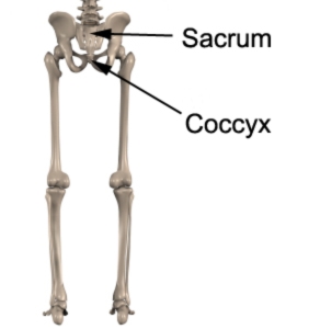
Figure 1 – Anatomy of the Sacrum and Coccyx
Sacrum
The sacrum is a heart shaped, or inverted triangular shaped bone, located at the base of the spine. The upper, or superior half, of the sacrum is wider and stronger, as it has an important role in distributing weight through to the pelvis via the strong sacroiliac joints. The lower, inferior half is non-weight bearing, and is therefore, narrower.
The sacrum has an opening, the sacral canal, through which travels the sacral plexus (nerves). The sacrum is composed of five fused sacral vertebrae (named S1 to S5 from top to bottom). Each of these fused vertebrae have a pair of foramina (holes) (posterior and anterior sacral foramina) out of which exit the sacral nerves.
Interestingly, the sacral vertebrae do not start to fuse together until about 16-18 years of age, with fusion normally completed by 34 years.
The front (ventral) surface of the sacrum is concave and smooth whereas the back (dorsal) surface is convex and covered by five rough longitudinal ridges.
The sacrum sits tipping forwards. This helps widen the opening of the lower part of the pelvis (known as the inferior pelvic aperture) and is particularly more pronounced in females to help accommodate child birth delivery.
Location
The sacrum is located at the bottom of the spine and is hugged on either side by the ilia of the pelvis. The fused sacral vertebrae are numbered 1-5 from top to bottom.
Forms Joints With
The lumbar spine – the ‘base’ of the sacrum, S1, forms joints with the L5 vertebrae via the facet joints and via an intervertebral disc. This is known as the lumbosacral joint (L5/S1). The iliolumbar ligaments assist in stabilizing this joint by attaching L5 to the sacrum and ilium of the pelvis.
The coccyx – the sacral coruna, found at the lower (inferior end) or ‘apex’ of the sacrum, forms a joint with (articulates with) the coccyx bone at the coccygeal coruna (known as the sacrococcygeal joint. This is a secondary cartilaginous joint in which ligaments and fibrocartilage both join the bone ends together. There is also a thin fibrocartilaginous disc in the joint. Movements at this joint are quite small, involving flexion and extension, however, they are quite important for childbirth. Occasionally, by an anatomical anomaly, this joint can be a synovial joint, subsequently allowing the joint to move more freely.
The Ilium of the pelvis – the left and right sacroiliac joints are formed between (the alar of) the sacrum and (the medial aspect of) the ilium of the pelvis. These strong synovial joints need to be very stable to allow for efficient distribution of load from the spine to the lower limbs (legs) and vice versa. Contributing to the stability of these sacroiliac joints are:
- irregular elevations and depressions in the bone surfaces (resulting in interlocking)
- a strong articular capsule
- the way the sacrum suspends like a wedge between the two iliac bones
- multiple ligamentous structures
The interosseus sacroiliac and the anterior and posterior sacroiliac ligaments provide primary stability, with secondary stability provided by accessory ligaments (iliolumbar, sacrospinous and sacrotuberous ligaments. Together, these ligaments resist ‘nutation’ and ‘counter-nutation’ movements at the sacroiliac joint.
Coccyx
The coccyx, or tail bone, is the remains of the embryonic ‘tail’ that exists until the 8th week of gestation. It serves as an attachment point for muscles, including gluteus maximus and the coccygeal muscles, and, the anococcygeal ligament. Unlike the rest of the vertebral column, it has no role in weight-bearing.
Like the sacrum, the coccyx is composed of vertebrae. Normally there are 4 coccygeal vertebrae, however, occasionally there may be 3 or 5. They are numbered from Co1 to Co4 (from top to bottom). Co1 is the largest. Its ‘horns’ (coccygeal coruna) form a joint (articulation) with the sacrum. Co2 to Co4 tend to fuse together during middle life, whilst Co1 tends to fuse with the sacrum during old age.
Muscles attachments
There are 35 muscles in total that attach to the sacrum, coccyx or pelvic bones. They include:
- Adductor brevis
- Adductor longus
- Adductor magnus
- Biceps femoris (long head)
- Coccygeus
- Erector spinae
- External oblique
- Gluteus maxiumus
- Gluteus medius
- Gluteus minimus
- Gracilis
- Iliacus
- Inferior gemellus
- Internal oblique
- Latissimus dorsi
- Levator ani
- Multifidus
- Obturator internus
- Obturator externus
- Pectineus
- Piriformis
- Psoas minor
- Pyramidalis
- Quadratus femoris
- Quadratus lumborum
- Rectus abdominis
- Rectus femoris
- Sartorius
- Semimembranosus
- Semitendonosus
- Sphincter urethrae
- Superficial transverse perineal ischiocavernous
- Superior gemellus
- Tensor fascia lata
- Transversus abdominus
Related injuries
- Sacroiliac Joint Dysfunction
- Piriformis Syndrome
- Gluteal Strain
- Lumbar Disc Bulge
- Facet Joint Sprain
- Sciatica
- Spinal Degeneration
- Coccydynia
Relevant Physiotherapy Exercises
- Lower Back Stretches
- Lower Back Strengthening Exercises
- Core Exercises
- Pilates Exercises
- Gluteal Strengthening Exercises
- Gluteal Stretches

Recommended Reading
- View our Lower Back Diagnosis Guide.
- View our Buttock Back Diagnosis Guide.
- View detailed information on improving your Posture.
- View detailed information on Postural Taping.
- View detailed information on Ergonomic Computer Setup.
- View detailed information on Choosing a School Bag.
- View detailed information on Safe Lifting.

Find a Physio
Find a physiotherapist in your local area who can diagnose and treat sports and spinal injuries and provide education on the anatomy of the sacrum and coccyx.

Link to this Page
If you would like to link to this article on your website, simply copy the code below and add it to your page:
<a href="https://physioadvisor.com.au/health/anatomy/bones/sacrum-and-coccyx”>Sacrum and Coccyx – PhysioAdvisor.com</a><br/>PhysioAdvisor provides detailed physiotherapy information on the human anatomy of the sacrum and coccyx. Including location, joints, muscular attachments, relevant injuries and more...
Return to the top of Sacrum and Coccyx.