Thoracic Vertebrae
Updated:
Relevant Bony Anatomy
The thoracic vertebrae (of the upper back) are larger than the cervical vertebrae (of the neck) and smaller than the lumbar vertebrae (of the lower back). There are twelve in total (T1 to T12), which together form the thoracic spine. They are numbered from the top of the thoracic spine (T1 – whose spinous process is the most prominent bony landmark just below the base of the neck) to the bottom of the thoracic spine (T12). The 1st, and 9th through to 12th thoracic vertebrae are atypical thoracic vertebrae with unique features (making them more similar to cervical and lumbar vertebrae respectively), whilst T2 to T8 are considered typical thoracic vertebrae and possess similar features.
Typical thoracic vertebrae primarily comprise of a cylindrical / heart shaped body (at the front of the bone) and a vertebral arch (situated directly behind the body), which forms a hole known as the vertebral foramen which, in the thoracic spine, is relatively smaller than it is in the cervical or lumbar regions. Since each vertebrae is situated directly above or below each other, their collective vertebral foramen line up forming the vertebral canal which houses and protects the spinal cord.
Each thoracic vertebrae joins with adjacent vertebrae primarily at the facet joints (located on each side of the spine, towards the back of the spine) and the discs of the thoracic spine (located centrally at the front of the spine) (figure 1). Movements between each adjacent vertebrae are relatively small, but when summated over the entire vertebral column allow considerable mobility.
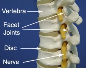
Figure 1 – Anatomy of the Thoracic Vertebrae
In the thoracic spine the facet joints are orientated in the coronal plane, which means the primary movements of the thoracic spine are axial rotation and lateral flexion (i.e. twisting and side bending of the spine). Towards the lower thoracic spine the facet joints progressively orientate more into the sagittal plane, similar to the facets of the lumbar spine. This orientation allows for greater flexion / extension movement (i.e. forward / backward bending of the spine) and less axial rotation / lateral flexion movement.
Thoracic vertebrae also have facets on either side of their bodies, which form joints with the heads of the corresponding ribs (known as the costovertebral joints). Each thoracic vertebrae also has various bony prominences, such as the spinous processes (located at the back of the bone) and transverse processes (located at each side of the vertebrae). These bony prominences provide attachment points to the ligaments and muscles of the thoracic spine. In the thoracic spine, the spinous processes are long and slender and orientated obliquely downwards, limiting the movement of spinal extension. The transverse processes also feature facets (except in the 11th and 12th thoracic vertebrae), which form joints with the corresponding ribs (known as the costotransverse joints). Together, the costovertebral and costotransverse joints, allow rotational movement of the ribs, which assists ventilation of the lungs.
Collectively the thoracic vertebrae act to support the head and the neck, protect the spinal cord, allow movement of the thoracic spine, ribs and thoracic cage, and provide attachment points for the muscles and ligaments of the thoracic spine.
Location
The twelve thoracic vertebrae are located in the mid section of the spine (upper back) between the neck and the lower back. They are numbered from the top to bottom – T1 to T12.
Forms Joints With
- C7 (lowest cervical vertebrae) – T1 forms joints with C7 via the facet joints and disc.
- Each of the vertebrae of the thoracic spine (T1 – T12) connect with the vertebrae above and below via bony processes on each side of the spine which form the facet joints and via the discs located centrally between each spinal segment.
- The ribs – at the costovertebral and costotransverse joints.
- L1 (first lumbar vertebrae) – T12 also forms joints with the first lumbar vertebrae (L1) via the facet joints and disc.
Major Muscles of the Thoracic Spine
Superficial Layer:
- Trapezius – this large back muscle attaches to the vertebral column from the base of the skull to the ligamentum nuchae of the neck, to the spinous processes of C7 and the thoracic spine down to T12. It connects the shoulder blade with the vertebral column and acts to support the arm and assist in upper limb movement by controlling shoulder blade movements.
- Latissimus Dorsi – this broad back muscle runs from the spinous processes of the lower thoracic vertebrae and lumbar spine, the connective tissue (fascia) of the thoracolumbar region, as well as the pelvis and lower ribs, to insert into the upper arm bone (humerus). Its primary action is to extend, adduct and internally rotate the upper arm (i.e. bring the upper arm backwards, to the side of the body and rotate the upper arm towards the body e.g. performing a chin up). It also influences movements of the shoulder blade and trunk.
- Rhomboids – these rhombus shaped muscles originate from the spinous processes of cervical and thoracic vertebrae (C7 to T5) and attach to the shoulder blade. The rhomboids main action is to pull the shoulder blades back (scapular retraction).
- Serratus Posterior Superior – is a thin, quadrilateral shaped muscle, located at the upper and back part of the thoracic spine. It lies deep to the rhomboids and aids inspiration by elevating ribs 2 to 5 where it attaches.
- Serratus Posterior Inferior – is located at the junction of the thoracic and lumbar regions. It originates from vertebrae T12 down to L3 and inserts into the 9th through to 12th ribs. It acts to draw the lower ribs backwards and downwards and assists in trunk rotation and extension. It also helps with forced expiration.
Intermediate Layer:
- Thoracic Erector Spinae – the main extensor (backward bending) muscle of the thoracic spine, located on either side of the vertebral column. Responsible for keeping the spine erect and helping to control forward and backward bending of the spine (flexion and extension). When acting unilaterally (on only one side of the body) it assists with side bending and spinal rotation to the same side.
Deep Layer:
- Transversospinalis Muscles – these shorter, deep muscles help stabilise the spinal segments. They are situated deep to the erector spinae, and run obliquely. They originate from transverse processes of inferior vertebrae and attach to spinous processes of superior vertebrae. Acting bilaterally (on both sides of the spine) these muscles produce segmental extension (backward bending of the spine) and stability. Acting unilaterally these muscles produce side flexion to the same side and rotation to the contralateral side (opposite side). These muscles (in order from superficial to deepest) are:
-
- Semispinalis
- Multifidus
- Rotatores
-
Other Attachments
- With the exception of T1, each thoracic vertebrae connects with the vertebrae above and below via an intervertebral disc.
- Numerous strong ligaments reinforce connections between adjacent vertebrae, discs and ribs, providing stability.
Related injuries
- Thoracic Disc Bulge
- Facet Joint Sprain
- T4 Syndrome
- Costovertebral Joint Sprain
- Scheuermann’s Disease
- Postural Syndrome
- Fractures

Relevant Physiotherapy Exercises

Recommended Reading
- View our Upper Back & Chest Diagnosis Guide.
- View detailed information on improving your Posture.
- View detailed information on Postural Taping.
- View detailed information on Ergonomic Computer Setup.
- View detailed information on Mobile Phone Ergonomics.
- View detailed information on Choosing a School Bag.
- View detailed information on Safe Lifting.

Find a Physio
Find a physiotherapist in your local area who can diagnose and treat sports and spinal injuries and provide education on the anatomy of the thoracic spine and vertebrae.

Link to this Page
If you would like to link to this article on your website, simply copy the code below and add it to your page:
<a href="https://physioadvisor.com.au/health/anatomy/bones/thoracic-vertebrae”>Thoracic Vertebrae – PhysioAdvisor.com</a><br/>PhysioAdvisor provides detailed physiotherapy information on the human anatomy of the thoracic spine and thoracic vertebrae. Including location, joints, muscular attachments, relevant injuries and more...
Return to the top of Thoracic Vertebrae.